


 Home
Home
 Products
Gastro Function
Pepsinogen I / Pepsinogen II (PGI/PGII)
Products
Gastro Function
Pepsinogen I / Pepsinogen II (PGI/PGII)
Pepsinogen I / Pepsinogen II (PGI/PGII)
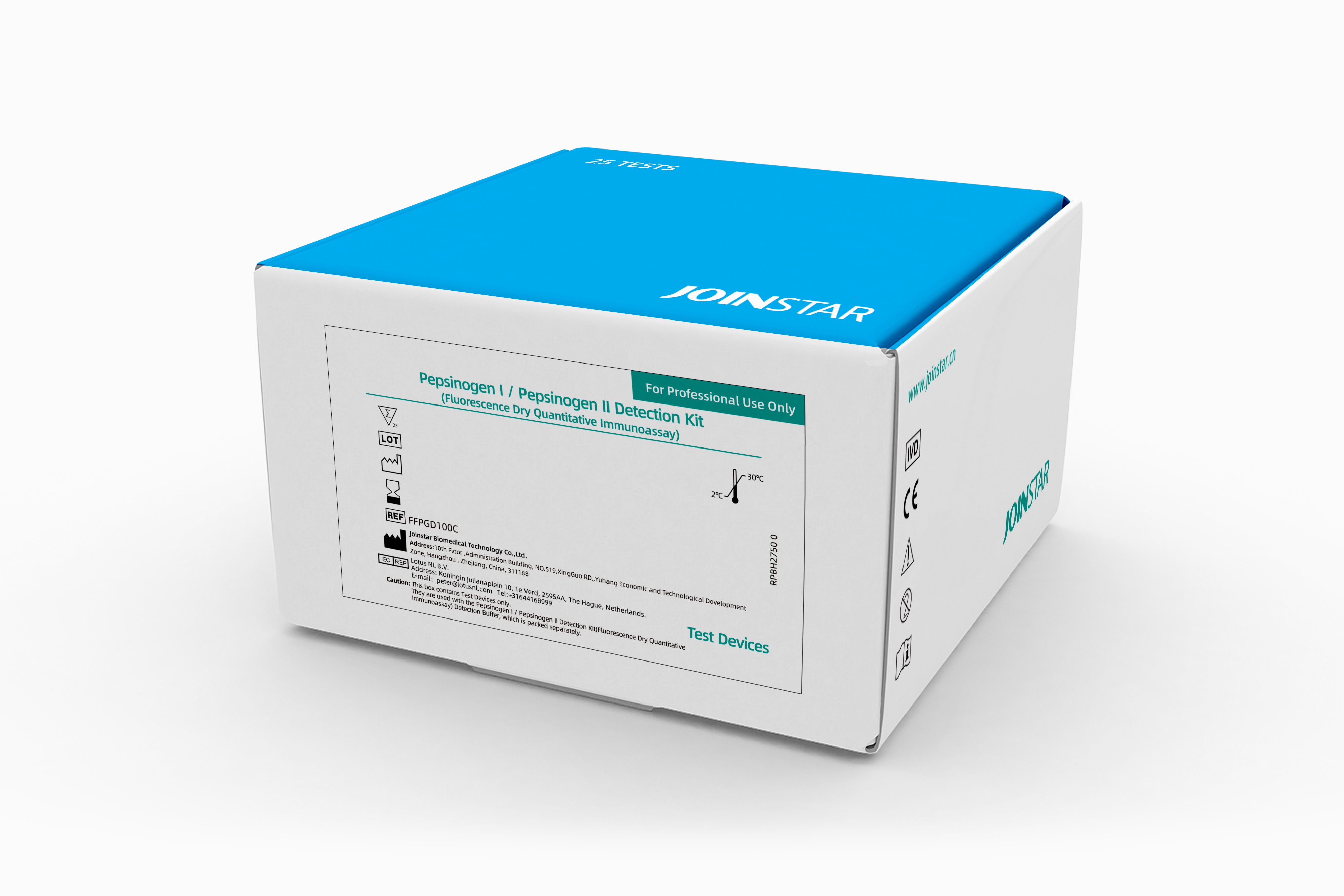
| Kit Name | PepsinogenI/PepsinogenII Detection Kit |
| Method | Fluorescence dry quantitative immunoassay |
| Assay Measuring Range |
PG I : 3.0 ng/mL ~ 200.0 ng/mL PG II : 1.5 ng/mL ~ 100.0 ng/mL |
| Incubation Time | 10 min |
| Sample Type | Human serum, Plasma (EDTA), Whole Blood (EDTA) |
| Reference Range |
PG I : > 70.0 ng/mL PG II : <27.0 ng/mL |
| Storage and Stability | Detection Buffer: Stable for up to 12 months (Store at 2°C–8°C in a sealed, dark environment). Reaction Plate: Stable for up to 12 months (Store sealed at 2°C–30°C). |
Order Information
| Catalog No. | Product | Test/Kit |
| FFPGD200 | PepsinogenI/PepsinogenII Detection Kit | 25 |

Introduction
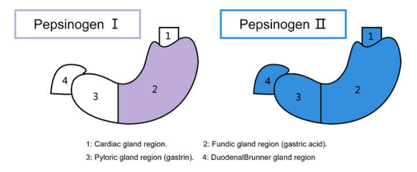
•Pepsinogen (PG) is the inactive precursor of pepsin and consists of 375 amino acids. Based on its biochemical properties, immunogenicity, cellular origin and intra-tissue distribution, PG can be divided into two subgroups, PG I and PG II. •PGⅠ is mainly secreted by the main cells and mucus neck cells of the fundic glands; PGⅡ is also produced by the main cells and mucus neck cells of the fundic glands, the mucus neck cells of the pyloric glands of the cardia and the gastric sinus, and the Brunner's gland in the upper duodenum, and a small amount of PGⅡ is also produced by the prostate and the pancreas. •Serum PGⅠ and PGⅡ reflect the number of gastric mucosal glands and cells, and also indirectly reflect the secretory function of different parts of the gastric mucosa. When pathological changes occur in the gastric mucosa, the serum PG level also changes. Since the stomach is almost the only source of PG, monitoring the concentration of PG in serum can be used as a means to monitor the status of the gastric mucosa.


Clinical Applications